Definition: The pleura is a closed serous sac, which is invaginated by the lung from its medial aspect.
Layers of the pleura:
The pleura is composed of two layers:

1 ) The outer layer (which lines the thoracic wall from inside) is known as the parietal pleura.

2) The inner layer (which covers the lung) is called the visceral pleura.

The visceral and parietal pleurae are continuous together around the root of the lung and the pleura extends
downwards as a fold
below the root called the pulmonary ligament.

The closed cavity between the visceral and parietal pleurae is called the pleural cavity.

Visceral pleura

• The inner layer of the pleura that Covers the surfaces of the lung and lines its fissures.

• It is absent in the hilum of the lung and between the two layers of the pulmonary ligament.

Parietal pleura

• Is the layer of the pleura that lines the chest wall and covers the mediastinum.

• Parts of the parietal pleura:

The parietal pleura is divided into four parts: costal, cervical, mediastinal and diaphragmatic.

1) Costal pleura:

It is the part of the parietal pleura lining the sternum, ribs, costal cartilages,
intercostal spaces and sides of the vertebral column.
It is continuous with the mediastinal pleura at the costomediastinal reflection and
with the diaphragmatic pleura at the
costodiaphragmatic reflection.

2) Cervical pleura:

It is the part of the parietal pleura, which covers the apex of the lung.

It is attached above by the suprapleural membrane.


Relations of the cervical pleura:
 The summit: is related to:

1 ) The costo-cervical trunk.

2) The suprapleural membrane.
 Anteriorly:

1 ) The subclavian artery: arched in front of it.

2) The scalenus anterior muscle.

3) The internal mammary artery.
 Posteriorly ( structures in front of the neck of the 1 st rib):

1 ) The stellate ganglion of the sympathetic trunk.

2) The superior intercostal artery.

3) The first thoracic nerve.
 Laterally:

1 ) The scalenus medius.

2) The scalenus posterior muscle.
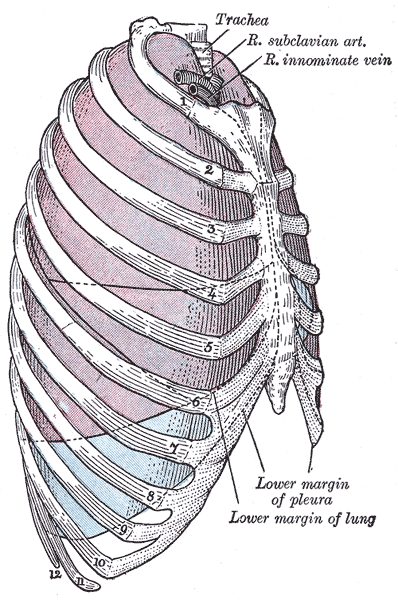
 Medially:

1 ) The trachea and the esophagus.

2) On the right side: the brachiocephalic artery and the right brachiocephalic vein.

3) On the left side: the left subclavian and left brachiocephalic vein.